Scientists found plastic in 100% of human brain samples they tested. Should we be concerned?
A team of scientists in New Mexico tested human brains and found microplastics in 100% of them. The worst part: the brains of people who died with dementia had the most plastic of all. In a separate study at Nanjing University, mice drank water with tiny plastic particles for 6 months, and 100% of the groups drinking plastic did worse on a memory test. Three more labs ran their own mouse studies. Every time, the mice given plastic did worse on a memory test. In mice, plastic in drinking water reached the brain and damaged memory, and in people, the brains of dementia patients held the most plastic of all.
6 minute read · Updated · By Aelo Research

Short Summary
A University of New Mexico team tested brain samples from about 90 people who had died. Every brain sample held microplastics, tiny flakes far thinner than a hair. The 12 brains from people who died with dementia held the most plastic of all. So what can plastic in the brain do? Labs have tested that in mice.[7]
At Nanjing University, male mice drank water with tiny plastic particles for 180 days. Scientists tagged the particles so they lit up under special light. After 6 months, the tagged plastic showed up in the mice's brains. In a swimming test, the mice had to remember where a hidden platform was. Every plastic group spent less time searching the right spot than the mice on clean water. The worst group spent less than half as long. That test measures learning and remembering, the skills a student leans on at exam time.[1]
Three more labs found the same: mice given plastic did worse on memory tests. In mice, plastic in drinking water reached the brain and damaged memory. In people, the brains of dementia patients held the most plastic of all. Below, we walk through each study: what the scientists did, what they found and why it matters to you.[1, 3, 4, 6, 7]
Do animal studies matter for humans?
Yes. These mice took in plastic one way people do: in the water they drank each day, for 6 months. A mouse brain has the same parts these teams measured: a memory center, a protective barrier and tiny blood vessels. The narrowest human vessels are about as narrow as a mouse's. And plastic has already turned up in human brains: in New Mexico, every one of about 90 brain samples tested held it, and the dementia brains held the most. The mice showed what plastic did. The human study showed it is there.[1, 6, 7]
Read our full article: Do animal studies matter for humans?
What did scientists find in human brains?
Plastic in every brain tested
In 2025, a New Mexico team tested brain, liver and kidney tissue from people who had died. Plastic turned up in all 3 organs, and in every one of about 90 brain samples. The bits in the brain were tiny flakes, far thinner than a hair. About 3 parts in 4 were polyethylene, the world's most common plastic.[7]
The 12 dementia brains held the most
The team also tested 12 brains from people who died with dementia. Those 12 held the most plastic of any group: about 5 times the amount in the other recent brains. The same table notes that the dementia values came down once more fat was removed, with no new number given.[7]
About 11 times more than the liver
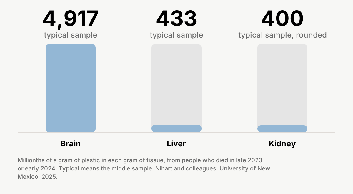
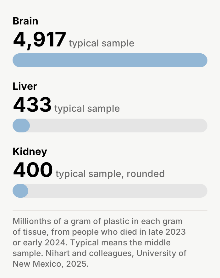
In the 2024 samples, a typical brain sample held about 11 times as much plastic as a typical liver sample, and the kidney held about as much as the liver.[7]
Which organ held the most plastic?[7]
At a glance: the Nanjing study
Did the plastic reach the mice's brains?
Found in 6 of 6 plastic groups
Yes. The particles were polystyrene, the plastic in foam cups. The team tagged them so they lit up under special light. Six groups drank particles in their water, in 3 sizes and 2 amounts; a seventh drank clean water. After 180 days, the tagged plastic showed up in the brains of all 6 plastic groups, and not in the clean-water mice.[1]
The brain's barrier let a dye through
The brain has a barrier that keeps most things in the blood out. In the plastic-water mice, a test dye got through it and reached the memory center. In clean-water mice, the dye stayed out.[1]
Did the mice do worse on memory tests?
6 of 6 plastic groups did worse
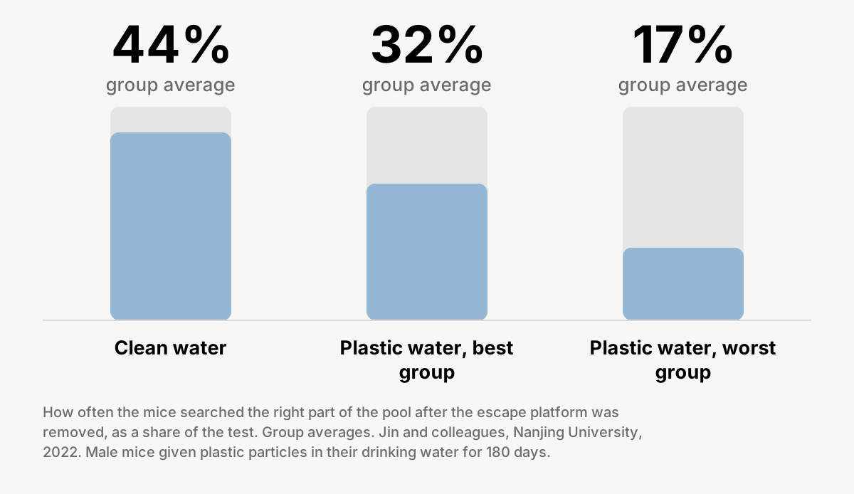
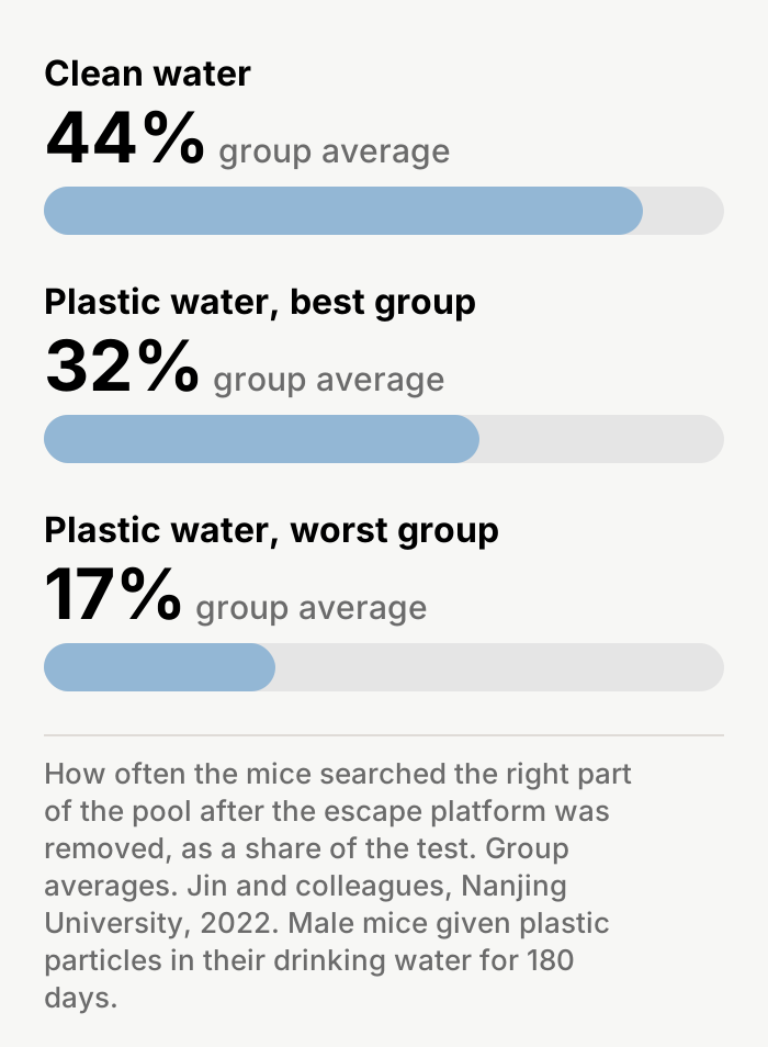
Yes. The test was a round pool with a small escape platform hidden just under the water. For 5 days, the mice learned where it was. On day 6, the team took the platform away and watched where each mouse went looking. Mice on clean water searched the right part of the pool 44% of the time. Mice in the 6 plastic groups searched it 17% to 32% of the time. Every plastic group did worse.[1]
Same speed, weaker memory
The plastic-water mice swam just as fast, and for the first 4 days they found the platform about as quickly. On a second memory test, with a new object to explore, all 6 plastic groups also scored lower, which the authors read as weaker short-term memory. In the mice on the higher dose, nerve cells in the memory center had about half as many connection points, or fewer, and that center showed more signs of inflammation.[1]
Which mice remembered where the platform was?[1]
Did other studies see the same thing in mice?
Three more studies, from three more labs
Yes. In Wuhan, mice got plastic by mouth every day for 4 weeks and did worse in the same kind of pool test. In Taiwan, mice got plastic for 8 weeks; it turned up in their memory center, and their memory got worse. A Beijing-led team put plastic straight into the blood of mice. Immune cells swallowed it, some of those cells got stuck in tiny brain vessels, and blood flow dropped. Those mice moved less and did worse on a maze memory test. When the same team fed 5 mice plastic through a tube, it showed up in brain blood vessels about 3 hours later, in 4 of the 5.[3, 4, 6]
What this proves, and what it doesn't
What these studies show
- Every one of about 90 human brain samples tested held plastic, and the 12 dementia brains held the most.[7]
- Brain samples from people who had died held about 11 times more plastic than liver samples.[7]
- Mice that drank plastic for 180 days had plastic in their brains.[1]
- Those mice did worse on the pool memory test, in all 6 plastic groups. Mice in 3 other labs also did worse on memory tests.[1, 3, 4, 6]
- In mice, cells full of plastic blocked tiny brain vessels and slowed blood flow.[6]
What these studies did not prove
- That plastic harms memory in people. The human study measured plastic, not memory.[7]
- That plastic causes dementia. The authors assume no cause, and dementia itself can raise the numbers.[7]
- The true amount in human brains. The test can read brain fat as plastic, 9 European scientists have challenged the method, and the dementia values came down after more fat was removed.[7, 9]
- Where the plastic in human brains came from. No water, food or air was measured.[7]
- That the mice got everyday amounts. The particles were new lab particles, and the Beijing-led tests used a shot into the blood.[1, 6]
Questions readers are asking
Were these studies done on people?
The 4 memory studies were done on mice. The New Mexico study tested tissue from about 90 people who had died. No study here tested memory in living people.[7]
About Aelo
Lab-tested on particles down to 0.5 microns (500 nanometers). Aelo's products are not connected to this research.
The technical record
Everything above is the story. Everything below is the paperwork: the exact statistics as published, the study’s own fine print, and the parts only a methods reviewer will want.
The numbers, exactly as published
| Plastic in every brain tested | Brain samples: 28 from 2016, 24 from 2024, 12 with dementia and 27 or 28 from East Coast brain banks (the paper's own counts conflict), about 90 in all. Every group's lowest value in Table S1 is above zero; "every sample" is this review's reading of that table, as the paper gives no per-sample count. Polyethylene 75% of brain plastic mass on average. |
|---|---|
| The 12 dementia brains held the most | 12 dementia cases: Alzheimer's disease 6, vascular dementia 3, other dementia 3, each confirmed by examining the brain after death. Median 26,076 µg/g from a single digestion step, against 4,917 in the 2024 brains (our arithmetic: 5.3 times); the table's footnote says later removal of fat with benzene "reduced values" and gives no reduced values. Higher than every normal group (two-sided t test, p < 0.0001). |
| About 11 times more than the liver | 2024 medians: brain 4,917 µg/g (middle half 4,026 to 5,608), liver 432.9 (main text: 433), kidney about 400. Brain against the other organs: two-way ANOVA p < 0.0001. The authors' own range: brain 7 to 30 times liver or kidney. |
| Found in 6 of 6 plastic groups | Whole-brain fluorescence imaging, 3 mice per group: signal in every plastic group and none in the control group (Figure 2; Figure S1). The method shows presence, not amount; the authors say it "cannot quantitatively detect the amount". |
| The brain's barrier let a dye through | Biotin tracer reached the hippocampus, hypothalamus and cortex in all 6 plastic groups and in none of the control mice; 3 mice per group, 3 slides each. |
| 6 of 6 plastic groups did worse | Day 6 probe trial, share of time in the target quadrant: control 44.37 ± 12.80%; plastic groups 16.87 ± 15.47% to 32.40 ± 15.68%; each plastic group against control p = 0.0013 to 0.0352 (one-way ANOVA, then Dunnett's test). |
| Same speed, weaker memory | Swim speed did not differ on any day (lowest p = 0.0512). Time to find the platform did not differ on days 1 to 4 (p = 0.10 to 1.00). Dendritic spines, 3 higher-dose groups: 4.50 to 6.50 per 10 µm against 12.25 in control (p = 0.0001 or lower); the number of mice counted is not stated. |
| Three more studies, from three more labs | Wang 2022: Kunming mice, 0.01, 0.1 or 1 mg a day by mouth for 4 weeks, saline control; abstract only. Lee 2022: 8 weeks; route not confirmed; abstract, highlights and supplement only. Huang 2025: male mice, a single injection (0.1 mg) or tube feeding (0.2 mg) of 5 µm particles; behavior tested after the injection; about 18% of plastic-filled cells stayed stuck until filming ended. |
The fine print: the Nanjing drinking-water study
Design: 7 groups of 12 male BALB/c mice, 6 weeks old at the start. Polystyrene particles 0.5, 4 or 10 micrometers across, at 100 or 1,000 micrograms per liter of drinking water, for 180 consecutive days. The authors estimate each mouse drank about 3 mL a day, or about 0.3 or 3 micrograms of plastic a day, based on the lab's earlier paper on the same batch of mice. Ten mice per group did the behavior tests; 3 per group were imaged for plastic in the brain, 3 had the dye test and 3 had electron microscopy.[1]
Barrier test: a tracer dye (biotin) put into the blood reached the hippocampus (memory center), hypothalamus and cortex in each of the 6 plastic groups and in none of the clean-water mice; 3 mice per group, 3 slides each. Electron microscope pictures of the 3 higher-dose groups showed shorter, thinner seals between the cells lining brain blood vessels.[1]
Plastic in the brain was shown by imaging the glow of whole brains. No amount was measured, and the authors write that the method "cannot quantitatively detect the amount". The brains were not described as flushed of blood, so the paper does not separate glow in the brain's blood from glow in brain tissue. No image shows plastic in the memory center specifically; what reached the memory center in the barrier test was a dye.[1]
Day 5 of practice: 2 of the 6 plastic groups (the higher dose of the 4 and 10 micrometer particles) took longer to find the platform, 46.81 and 54.72 seconds against 25.40 for clean water (p = 0.0457 and 0.0254). Days 1 to 4 did not differ.[1]
Platform crossings on day 6: the plastic-water mice crossed the platform's old spot a third as often as the clean-water mice, or less. Our check: those averages do not fit the paper's stated group size, so this review reports that result only as a ratio and prints no group size beside it.[1]
New-object test: the clean-water mice averaged 0.46, below the 0.5 the authors call the "no-preference score"; the plastic groups scored 0.22 to 0.27 (p = 0.0046 or lower). The authors read the lower score as weaker short-term memory and note the short 2-minute test and the mice's older age at testing. This article does not lead with that test.[1]
Nerve connections: in the 3 higher-dose groups, 4.50 to 6.50 connection points (dendritic spines) per 10 micrometers of nerve branch, against 12.25 in clean-water mice (p = 0.0001 or lower). The number of mice counted is not stated. Inflammation genes were more active in the memory center of plastic groups, measured in 3 mice per group.[1]
Erratum (PubMed 36512431, 13 December 2022): corrected the author affiliations and one abstract sentence from "higher" to "lower" dendritic spine density. The PMC full text carries the fix; the PubMed abstract still shows "higher".[2]
Loose ends we found: the text calls one group's day-5 swim path "significantly lower", while its own table shows it was longer (377.74 mm against 184.70 mm); several gene-test p-values do not track the size of the differences; blinding is not stated.[1]
Limits the authors state: the particles were new lab particles, not worn environmental plastic; the method shows plastic reaching the brain but not how much; the 3 particle sizes did not make a clear difference. Our notes: the authors describe their doses as "environmental pollution concentrations", citing a review of marine pollution rather than drinking-water measurements. The same lab's testes paper used mice from the same batch, so it is not an independent repeat.[1]
Funding: National Natural Science Foundation of China (31870492, 31901182, 31971517); Natural Science Foundation of Jiangsu Province (BK20190316); Fundamental Research Funds for the Central Universities (0214-14380438, 0214-14380471). The authors declare no competing financial interests.[1]
The fine print: the Wuhan, Taiwan and Beijing-led studies
Wuhan (Wang 2022, Central China Normal University): only the abstract, the supplement's particle-check figure and the graphical abstract could be read. The full text is paywalled, and no legal open copy was found. The route is given only as "orally exposed", with 0.01, 0.1 or 1 mg a day for 4 weeks and saline for the comparison mice. Not yet known from the paper: group sizes, sex and age of the mice, particle size, how the plastic was given by mouth, every maze number, which doses had an effect, where acetylcholine (the messenger chemical) was measured, and the vitamin E dose and timing. Vitamin E was not tested in people, and this article gives no supplement advice. Reviews that describe the study disagree on its length and particle size, so none of their numbers are used here. The title states a cause ("by inducing oxidative stress and decreasing the level of acetylcholine"); that is the authors' conclusion. Funding and conflicts of interest are in the unread full text.[3]
Taiwan (Lee 2022): read were the abstract, the 4 highlights, the 7 figures (without their legends), the supplement and the correction. The paper is open access, but the publisher's site blocked automated readers, so the Methods and Results text were not read. The route, dose, particle size, mouse strain and age, and behavior group sizes are therefore not confirmed here. Two later papers that cite it describe a feeding tube; that is not used as fact. From the supplement's own table: memory-center synapse proteins, 5 mice per group, Arc lower at 8 weeks (p = 0.029) and GluA1 higher (p = 0.022). Corrigendum (PubMed 37088020, 2023): added the missing swim-path images to Figure 7H and corrected Chang Gung grant numbers; no finding changed. Funding: Ministry of Science and Technology, Taiwan (110-2320-B-008-002, 109-2314-B-038-078-MY3, 2320-B-008) and Chang Gung Memorial Hospital (CMRPF6H0051, 52, 53; CMRPF6L0081, 0082). The competing-interest statement is in the unread body text.[4, 5]
Beijing-led (Huang 2025, Chinese Research Academy of Environmental Sciences and Peking University): the feeding-tube arm got one dose of 100 microliters at 2 mg/ml (0.2 mg of plastic) and was imaged from 1 hour to about 3 hours 40 minutes after dosing; first flash at 2 hours 20 minutes, median 191 minutes, 23 flashes across 4 of 5 mice. The injection arm got 100 microliters at 1 mg/ml (0.1 mg), which the authors estimate gives about 50 micrograms per milliliter of blood; lower-dose arms gave about 25 and 5. Particles were 5 micrometers across, with 2 and 0.08 micrometer particles in a size test. About 94% of the plastic-carrying cells were immune cells. Blood flow in the smaller brain vessels fell within 10 minutes and hit its low point at 30 minutes. Some stuck cells were still there 7 days later; fewer again at 28 days, but not zero. Behavior: open field 10 to 11 mice per group, Y-maze 11, spinning rod and hanging test 12, against saline-injected controls; the hanging test was normal by day 7 and the spinning rod by day 28. The authors write that the plastic's effect on movement "may influence performance in open field and Y-maze tests". Method notes: the experimenters knew which mice got plastic; many imaging results count fields of view rather than mice; Student's t tests with no correction for multiple comparisons; the blood-flow test ran under anesthesia, with no saline comparison and no stated number of mice. The title's "cerebral thrombosis" goes beyond what was measured: no clot parts were imaged, and the authors say platelet imaging is still needed. They also write: "It is premature to directly apply this mechanism to human research systems." Funding: Fundamental Research Funds for the Central Public-interest Scientific Institution (2022YSKY-34) and the National Natural Science Foundation of China (51908524); no competing interests declared.[6]
The fine print: the New Mexico study
Medians, in micrograms per gram of wet tissue: brain 3,345 in 2016 (middle half 1,267 to 5,213) and 4,917 in 2024 (4,026 to 5,608); liver 103.7 and 432.9; kidney about 400 in both years (the main text and the supplement give the 2024 kidney figure with different rounding, so this review prints it as about 400). East Coast brains, 1997 to 2013: 1,254. Our arithmetic on the 2024 medians: brain 11.4 times liver and 12.1 times kidney, printed as "about 11 times". The authors' own summary: 7 to 30 times.[7]
Dementia brains: 12, median 26,076 (middle half 17,629 to 35,831). The table's footnote says these values came from a single digestion step and that later removal of fat with benzene reduced them; no reduced values are given.[7]
Group sizes: 28 brains from 2016, 24 from 2024 and 12 with dementia. The East Coast count conflicts inside the paper (28 in the Methods, 27 in Table S1), so this review says about 90 brains in all. No sample-size calculation was made, and the dementia and East Coast groups were added later, in batches, after peer reviewers asked for them.[7]
Time trend: the abstract gives p = 0.01 for 2016 against 2024. In the fullest model, adjusting for age, sex, race, ethnicity and cause of death, the date effect was 0.17 (95% CI −0.06 to 0.39, p = 0.14); in a date-only model, 0.24 (0.05 to 0.43, p = 0.02). Age showed no link with brain plastic (p = 0.87). The oldest brains came from the East Coast, so place and year are mixed.[7]
Polyethylene made up 75% of the plastic mass in brain on average. The main text says infrared checks on 5 brains confirmed this, but in Supplementary Table 14 no polyethylene match reached the paper's own 60% threshold (best 53.88%).[7]
The test dissolves the tissue, heats a speck of what is left into gas and reads the chemical fingerprint, the same method as the Italian artery study. The digestion left more behind in brain: it dissolved 99.4% of liver and kidney mass but 91.8% of brain, leaving pellets of about 3 mg against 41 mg from 500 mg of tissue. The authors say this tracks the plastic measured; they also note that lipids can interfere with the polyethylene reading. The brain is the fattiest organ tested.[7]
Checks: a second lab, at Oklahoma State University, tested 5 brains from 2016 and matched (p = 0.49); repeat readings of one sample varied by about 25%. Blanks covered the digestion liquids, tubes and pipette tips, but not the polyethylene cutting boards, stock jars or brain buckets, which the authors call "the only known potential exposure to exogenous plastic". The 2016 samples sat in plastic jars for 7 to 8 years and the 2024 samples for 2 to 4 months, yet the newer ones held more, so the jars cannot explain the rise, the team says. A blank filter held 25 particles in a dye count, against 63 to 502 in brain samples (our reading of Table S12).[7]
Author Correction (PubMed 40164728, 31 March 2025): removed duplicated supplementary images, fixed scale bars, added an ethanol step to the method and clarified Supplementary Table 12. It states the fixes "do not affect spectra data found in Table S14".[8]
Challenge (PubMed 41233597), published in the same journal in November 2025: 9 authors from the University of Padua, UFZ Leipzig, the University of Birmingham, Potsdam University and IGB Berlin, BAM Berlin (a materials-testing institute), Utrecht University and Leiden University. Only its first paragraph could be read; it cites "limited contamination controls and lack of validation steps". The reply (PubMed 41233598), in the same issue, was also read to its first paragraph, which names the digestion method, possible interference from phospholipids (a kind of fat) and quality-control steps.[9, 10]
The senior author's slides for the Biomonitoring California panel, March 2026, are a talk, not a paper: "Lipids look like polyethylene", and "More aggressive digestion on a new cohort of ADRD samples reveals lower concentrations but consistent trends." The new numbers appear only on chart images.[11]
The authors write that their data "are associative and do not establish a causal role". Funding: US National Institutes of Health (P20 GM130422, K12 GM088021, R01 ES014639, R01 ES032037, P30 ES032755, R15 ES034901, P50 MD015706, UL1 TR001449); the publisher's funder record also lists the PhRMA Foundation. The authors declare no competing interests.[7]
The fine print: how we checked this article
Read in full: the Nanjing study and its 47-page supplement, the Beijing-led study and its supplement, and the New Mexico study and its supplement. Read at abstract level: the Wuhan study (paywalled) and the Taiwan study (open access, but blocked to automated readers). The challenge and reply letters were read to their first paragraphs. No paywall or bot check was bypassed. The key numbers were re-checked against the PubMed records on 2026-10-05.[1, 3, 4, 6, 7]
The fine print: about this version
This version was shortened and rewritten in plainer words, with question headings. The standfirst and Short Summary now lead with what the New Mexico team found in human brains, including the dementia brains, then what happened to the mice and what three more studies found. The brain-against-liver comparison and the challenge to the measuring method appear in the body, with the organ chart. The section on whether animal studies matter for humans comes first, and the three smaller mouse studies share one section. To keep the page under its reading-time limit, three reader questions (whether the mice drank their plastic, whether brain plastic is rising, and how much plastic was in the water), the short section on the route from water to brain, the Beijing-led team's movement caveat and the lead scientist's later talk appear only in this record; the route facts also appear in the mouse sections above. The chart of brain plastic by year left the story, because the rise depends on the statistical model; its numbers are in this record. Every figure that left the story is printed in these sections, from the papers' own text and tables. Aelo does not turn a mouse dose into a human one. No figure changed.
Sources
- Jin H, Yang C, Jiang C, Li L, Pan M, Li D, Han X, Ding J. Evaluation of Neurotoxicity in BALB/c Mice following Chronic Exposure to Polystyrene Microplastics. Environ Health Perspect. 2022;130(10):107002. doi:10.1289/EHP10255. PMID 36251724. PMCID PMC9555296. Evaluation of Neurotoxicity in BALB/c Mice following Chronic Exposure to Polystyrene Microplastics
- Erratum. Environ Health Perspect. 2022;130(12):129001. doi:10.1289/EHP12418. PMID 36512431. Erratum: "Evaluation of Neurotoxicity in BALB/c Mice following Chronic Exposure to Polystyrene Microplastics"
- Wang S, Han Q, Wei Z, Wang Y, Xie J, Chen M. Polystyrene microplastics affect learning and memory in mice by inducing oxidative stress and decreasing the level of acetylcholine. Food Chem Toxicol. 2022;162:112904. doi:10.1016/j.fct.2022.112904. PMID 35257813. Abstract only. Polystyrene microplastics affect learning and memory in mice by inducing oxidative stress and decreasing the level of acetylcholine
- Lee CW, Hsu LF, Wu IL, Wang YL, Chen WC, Liu YJ, Yang LT, Tan CL, Luo YH, Wang CC, Chiu HW, Yang TC, Lin YY, Chang HA, Chiang YC, Chen CH, Lee MH, Peng KT, Huang CC. Exposure to polystyrene microplastics impairs hippocampus-dependent learning and memory in mice. J Hazard Mater. 2022;430:128431. doi:10.1016/j.jhazmat.2022.128431. PMID 35150991. Abstract, highlights and supplement. Exposure to polystyrene microplastics impairs hippocampus-dependent learning and memory in mice
- Corrigendum. J Hazard Mater. 2023;453:131398. doi:10.1016/j.jhazmat.2023.131398. PMID 37088020. Corrigendum to: "Exposure to polystyrene microplastics impairs hippocampus-dependent learning and memory in mice" J. Hazard. Mater. 430 (2022) 128431
- Huang H, Hou J, Li M, Wei F, Liao Y, Xi B. Microplastics in the bloodstream can induce cerebral thrombosis by causing cell obstruction and lead to neurobehavioral abnormalities. Sci Adv. 2025;11(4):eadr8243. doi:10.1126/sciadv.adr8243. PMID 39841831. PMCID PMC11753373. Microplastics in the bloodstream can induce cerebral thrombosis by causing cell obstruction and lead to neurobehavioral abnormalities
- Nihart AJ, Garcia MA, El Hayek E, Liu R, Olewine M, Kingston JD, Castillo EF, Gullapalli RR, Howard T, Bleske B, Scott J, Gonzalez-Estrella J, Gross JM, Spilde M, Adolphi NL, Gallego DF, Jarrell HS, Dvorscak G, Zuluaga-Ruiz ME, West AB, Campen MJ. Bioaccumulation of microplastics in decedent human brains. Nat Med. 2025;31(4):1114-1119. doi:10.1038/s41591-024-03453-1. PMID 39901044. PMCID PMC12003191. Bioaccumulation of microplastics in decedent human brains
- Author Correction. Nat Med. 2025;31:1367. doi:10.1038/s41591-025-03675-x. PMID 40164728. Author Correction: Bioaccumulation of microplastics in decedent human brains
- Monikh FA, Materić D, Valsami-Jones E, Grossart HP, Altmann K, Holzinger R, Lynch I, Stubenrauch J, Peijnenburg W. Challenges in studying microplastics in human brain. Nat Med. 2025;31(12):4034-4035. doi:10.1038/s41591-025-04045-3. PMID 41233597. First paragraph read. Challenges in studying microplastics in human brain
- Campen MJ, West AB, Garcia M, Gullapalli R, El Hayek E. Reply to: Challenges in studying microplastics in human brain. Nat Med. 2025;31(12):4036-4037. doi:10.1038/s41591-025-04046-2. PMID 41233598. First paragraph read. Reply to: Challenges in studying microplastics in human brain
- Campen M. Micro- and Nanoplastics and Human Health: Essential Key Studies. Slides for the Biomonitoring California Scientific Guidance Panel, March 2026. A talk, not a peer-reviewed paper. Micro- and Nanoplastics and Human Health: Essential Key Studies